The Missouri campus library has many anatomy and dental models available for use. Models must stay in the library and are not allowed in other parts of the campus.
* Per special request, models may be checked out for special events or by faculty.

Model Type: Skeleton
Model Features:

Model Type: Skeleton
Model Features:

Model Type: Skeleton
Model Features:

Model Type: Skeleton
Model Features:

Model Type: Skeleton
Model Features:

Model Type: Box of bones
Model Features:

Model Type: Spine
Model Features:

Model Type: Spine
Model Features:

Model Type: Spine
Model Features:
*By Special Request Only: Please see 3D Print Shop manager for more information
Patent Number: 11798433

Model Type: Spine
Model Features:

Model Type: Spine
Model Features:

Model Type: Spine
Model Features:

Model Type: Spine
Model Features:

Model Type: Spine
Model Features:


Model Type: Spine
Model Features:

Model Type: Spine
Model Features:

Model Type: Spine
Model Features:
![]()

Model Type: Cranial/Cervical
Model Features:

Model Type: Cranial

Model Type: Cranial

Model Type: Cranial


Model Type: Skull
Model Features:

Model Type: Skull
Model Features:

Model Type: Skull
Model Features:

Model Type: Skull
Model Features:

Model Type: Skull
Model Features:

Model Type: Skull
Model Features:

Model Type: Skull
Model Features:

Model Type: Skull
Model Features:

Model Type: Skull
Model Features:

Model Type: Organ
Model Features:

Model Type: Organ
Model Features:

Model Type: Organ
Model Features:

Model Type: Organ
Model Features:

Model Type: Organ
Model Features:

Model Type: Organ
Model Features:

Model Type: Organ
Model Features:

Model Type: Organ
Model Features:

Model Type: Organ
Model Features:

Model Type: Organ
Model Features: The 3B Scientific Standard Pregnancy Series with 5 Models shows the most important stages of development of embryo or fetus.

Model Type: Organ
Model Features:
Model Type: Chest
Model Features:

Model Type: Chest
Model Features:

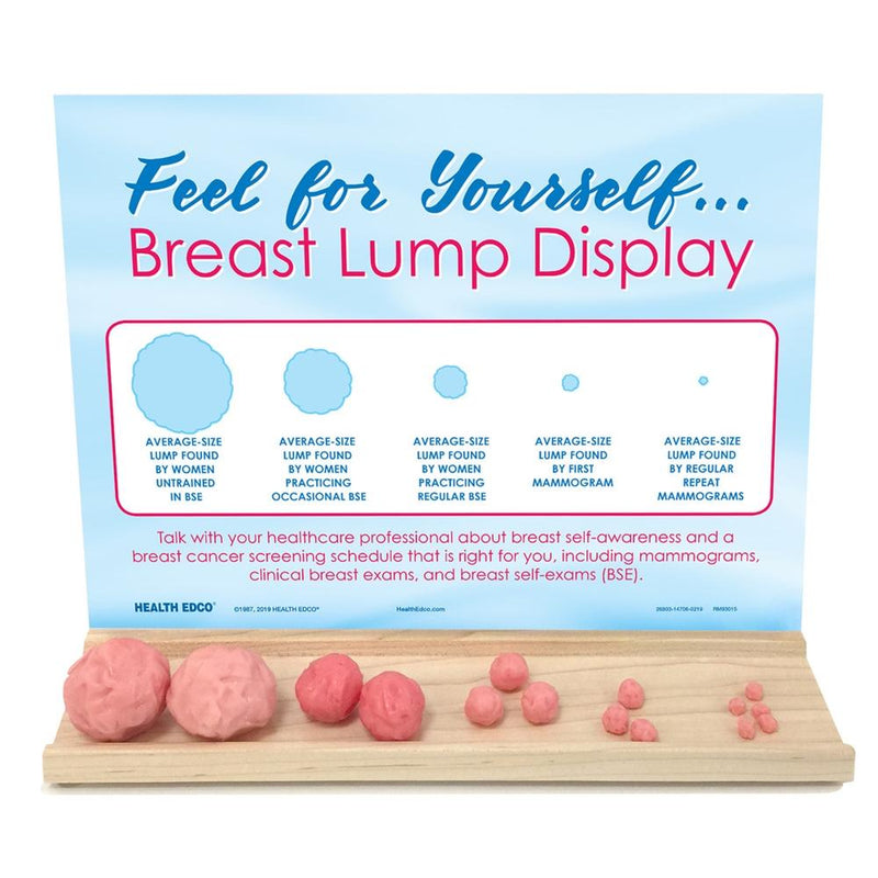
Model Type: Reproduction
Model Features:

Model Type: Reproduction
Model Features:

Model Type: Reproduction
Model Features:

Model Type: Reproduction
Mode Features:

Model Type: Reproduction
Model Features:

Model Type: Reproduction
Model Features:

Model Type: Reproduction
Model Features:

Model Type: Reproduction
Model Features:

Model Type: Reproduction
Model Features:

Model Type: Reproduction
Model Features:
*By Special Request Only: Please see 3D Print Shop manager for more information
Patent number: 11798433

Model Type: Upper Chest
Model Features:

Model Type: Organ, Skin
Model Features:

Model Type: Audiology
Model Features:

Model Type: Audiology
Model Features:

Model Type: Audiology
Model Features:

Model Features:

Model Type: Audiology
Model Features:

Model Type: Brain
Model Features:

Model Type: Brain
Model Features:

Model Type: Brain
Model Features:

Model Type: Brain
Model Features:

Model Type: Head
Model Features:

Model Type: Head
Model Features:

Mode Type: Head
Model Features:

Model Type: Head
Model Features:

Model Type: Head and Neck
Model Features:

Model Type: Larynx
Model Features:

Model Type: Larynx
Model Features:

Model Type: Larynx
Model Features:

Model Type: Tongue and Larynx
Model Features:

Model Type: Head
Model Features:

Model Type: Head
Model Features:

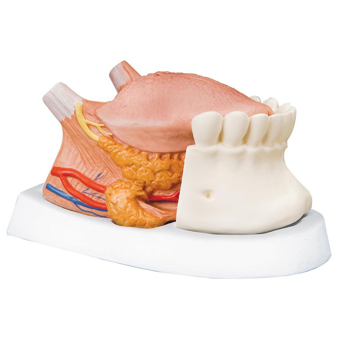
Model Type: Dental
Model Features:

Model Type: Dental
Model Features:

Model Type: Dental
Model Features:

Model Type: Dental
Model Features:

Model Type: Dental
Model Features:

Model Type: Dental
Model Features:

Model Type: Dental
Model Features:

Model Type: Dental
Model Features:

Model Type: Dental
Model Features:

Model Types: Dental
Model Features:

Model Type: Dental
Model Features:
Model Type: Dental
Model Features:

Model Type: Dental
Model Features:

Model Type: Dental
Model Features:

Model Type: Dental
Model Features:

Model Type: Dental, Skull
Model Features:

Model Type: Foot
Model Features:

Model Type: Foot
Model Features:

Model Type: Lower limb
Model Features:


Model Type: Foot
Model Features:

Model Type: Leg
Model Features:

Model Type: Leg
Model Features:

Model Type: Leg
Model Features:

Model Type: Leg
Model Features:

Model Type: Lower Limb
Model Features:

Model Type: Lower Limb
Model Features:

Model Type: Arm
Model Features:

Model Type: Arm
Model Features:

Model Type: Arm
Model Features:

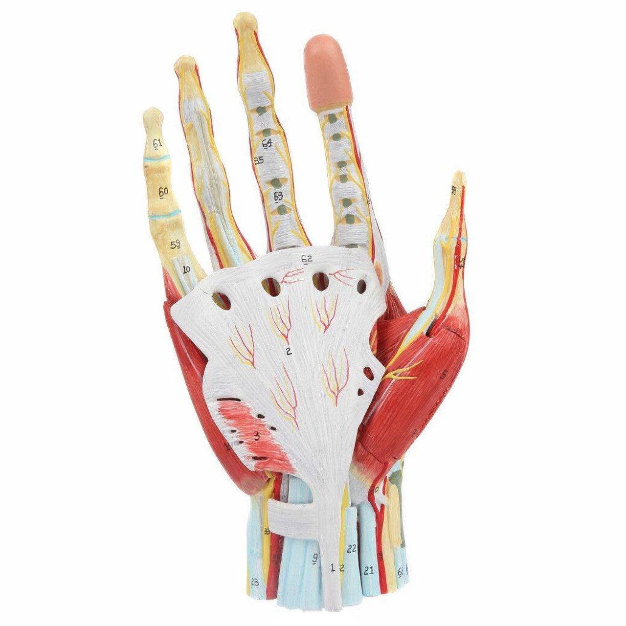
Model Type: Hand
Model Features:

Model Type: Hand
Model Features:

Model Type: Hand
Model Features:
Model Type: Hand
Model Features:

Model Type: Arm
Model Features:

Shoulder and Cervical Vertebrae
Model Type: Shoulder
Model Features:
We have received permission as a courtesy to use the pictures of each model from Anatomy Warehouse and Bone Clone.

Model Type: Upper Chest
Model Features:
Model Type: Arm
Model Features:
Model Type: Arm
Model Features:
Model Type: Leg
Model Features:
Model Type: Skeleton
Model Features:
Model Type: Organ
Model Features: